We are fortunate at Vista Eyecare to have doctors who maintain a high standard of equipping our office with cutting-edge technology. Our doctors of optometry want to provide their patients with the best imaging and diagnostic tools available to impart the highest standard of ocular care.
Medmont Automated Perimeter
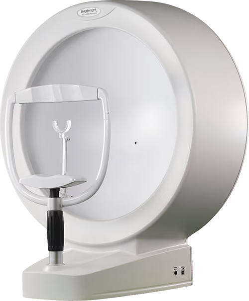
The Medmont Automated Perimeter performs screening and threshold tests of visual fields. The clinical applications of visual field testing include tests for glaucoma, flicker perimetry, binocular testing (including driving tests), neurological testing, macula testing, peripheral vision testing, and others.
IDRA Device
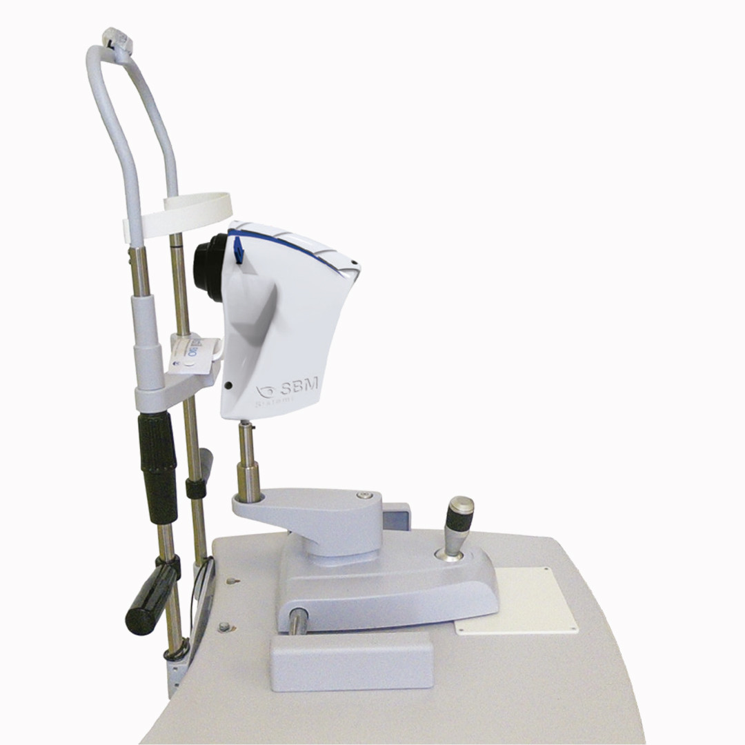
The IDRA device is a comprehensive dry eye testing system that is used for both screening patients as well as performing extensive dry eye assessments. Using this device, we can perform meibography, which refers to a non-invasive eye test that assesses the health of a patient’s meibomian glands. Meibography is often done to assess patients struggling with dry eye syndrome. Idra is capable of analyzing a patient’s tear film including lipid content, tear stability, and blink patterns.
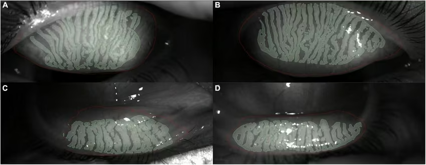
These are images taken by the infrared non-contact camera showing the meibomian gland pattern on the upper and lower eyelids. The software is then able to create a simulated 3D meibomian gland pattern. Images taken from Sanchez-Gonzalez et al., (2022).
Nidek TONOREF II Autorefractor Keratometer
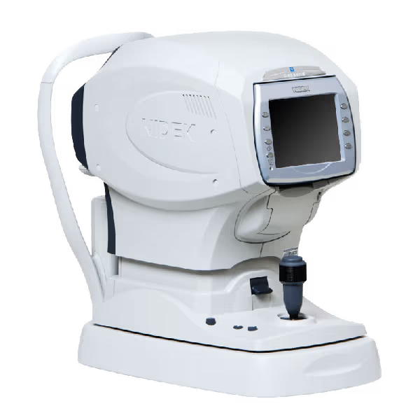
The Nidek TONOREF II Autorefractor Keratometer is designed to perform objective refraction, corneal shape measurement, and non-contact tonometry. Autorefraction is a scan taken of the curvature of the eye used to estimate prescription based on the patient’s refractive error. Tonometry is an eye test that measures intraocular pressure by providing several puffs of air to the surface of the eye. Measuring intraocular pressure is crucial for diagnosing eye diseases including glaucoma.
Optical Coherence Tomography
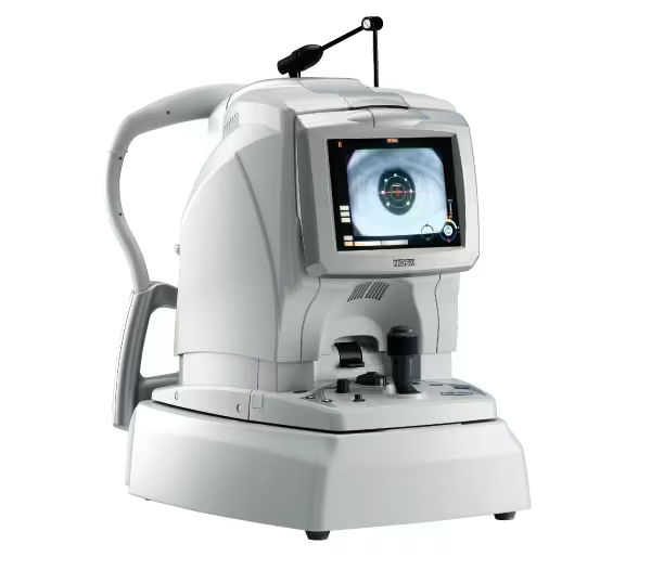
Optical Coherence Tomography (OCT) is an eye test that uses light waves to create detailed images of interior structures of the eye, including the optic nerve and retina. OCT is an important test to detect several eye diseases as well as provide early diagnosis and treatment.
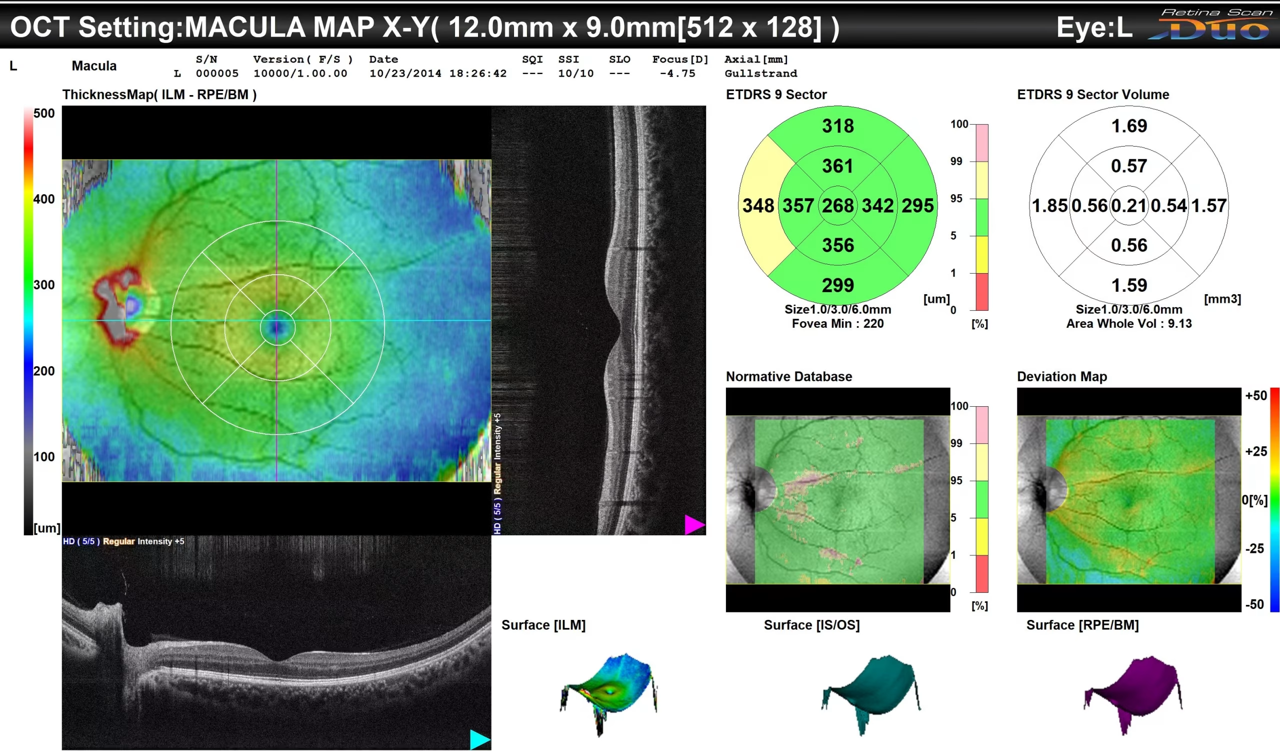
This is a macula map scan taken with Optical Coherence Tomography. This type of scan on the OCT provides high resolution images to analyze the thickness and structure of the different layers of the macula.
Optos
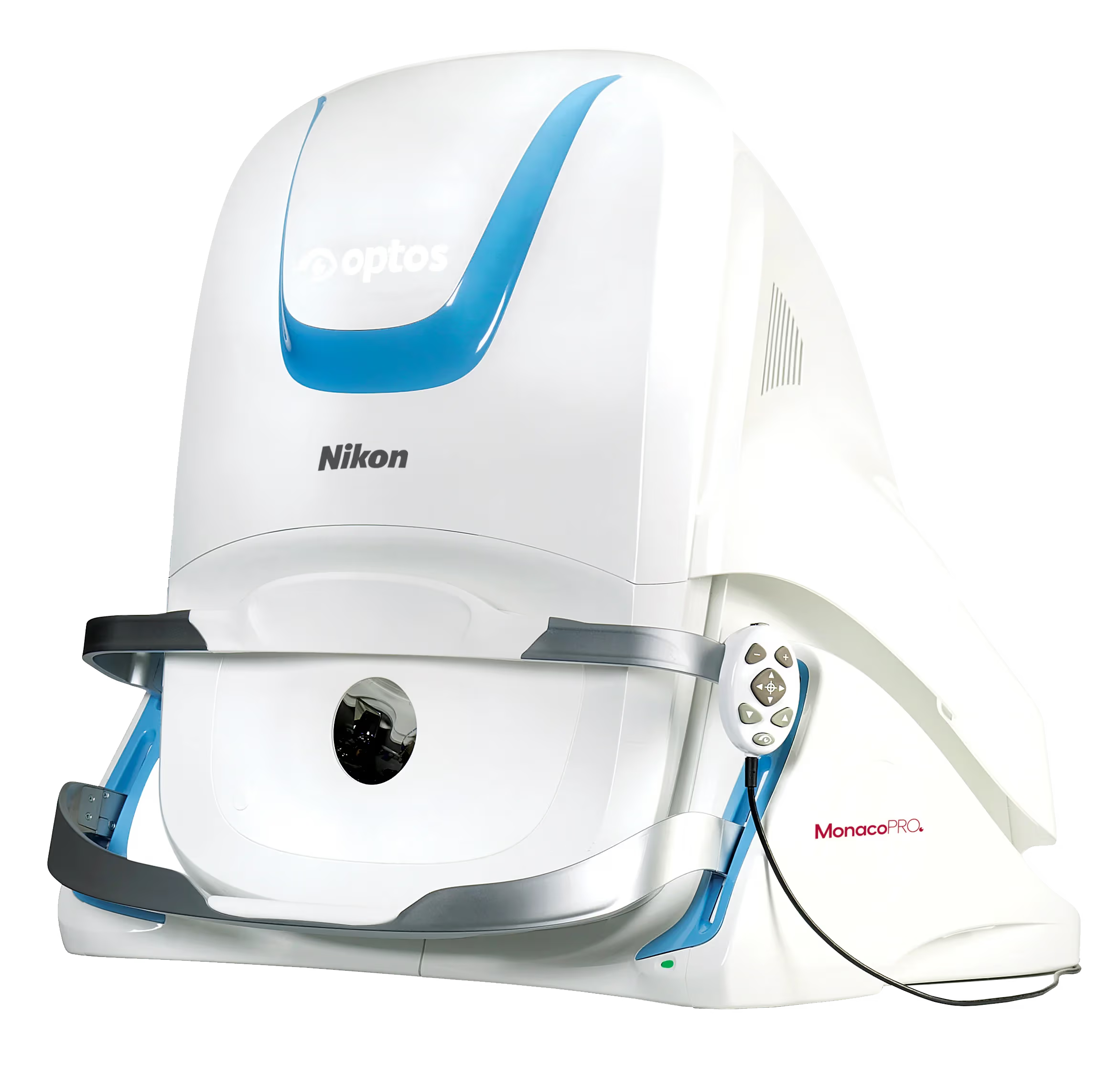
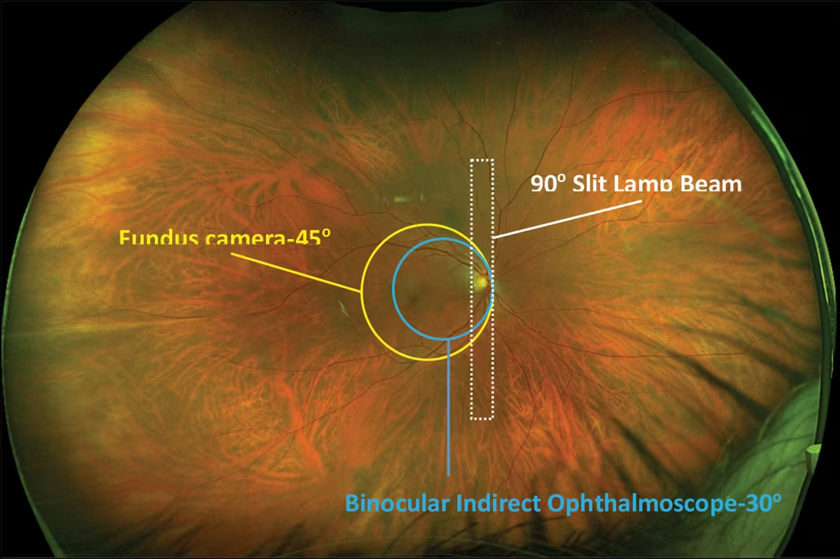
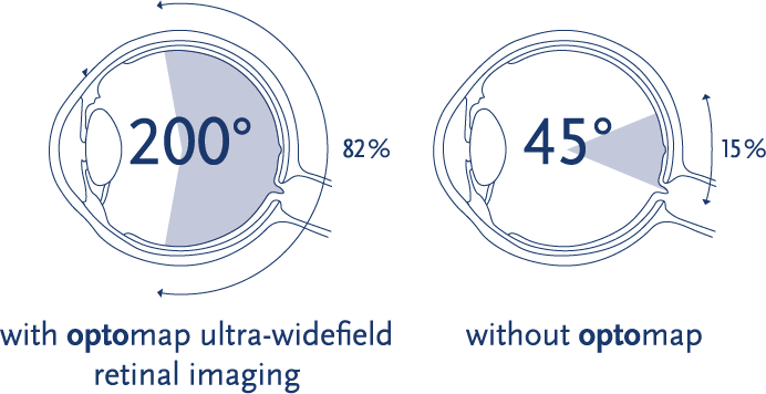
Optos ultra-widefield retinal imaging, or optomap imaging, is a non-invasive retinal imaging procedure that takes high-resolution images and scans of the retina. One of the newest machines in our office, this imaging provides the doctor with information to diagnose and treat various systemic and eye diseases.
Retinalogik
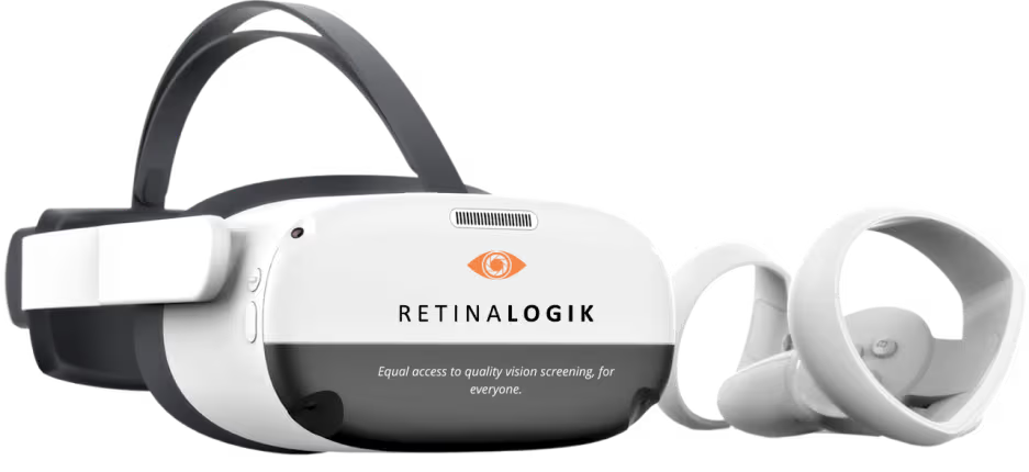
Using innovative virtual reality headsets, Retinalogik is a Canadian-based software company that is used to conduct various vision tests including visual field testing. This ophthalmic device is used to screen, monitor, and assist in the detection and progression monitoring of visual field defects.
References
Optos. (2023, November 20). Invest in your practice with optomap today. Optos. https://www.optos.com/blog/2023/november/invest-in-your-practice-with-optomap/
Sánchez-González, M. C., Capote-Puente, R., García-Romera, M.-C., De-Hita-Cantalejo, C., Bautista-Llamas, M.-J., Silva-Viguera, C., & Sánchez-González, J.-M. (2022). Dry eye disease and tear film assessment through a novel non-invasive ocular surface analyzer: The OSA protocol. Frontiers in Medicine, 9. https://doi.org/10.3389/fmed.2022.938484
Torbit, J., & Sutton, B. (2023, September 15). Ultra-widefield imaging: Expand your horizons. Review of Optometry. https://www.reviewofoptometry.com/article/ultrawidefield-imaging-expand-your-horizons
